
Chapter 1
Understanding your Diagnosis
| CHAPTER 1
Understand Your Diagnosis |
Where and What is the Prostate?
The prostate is a walnut-sized gland that makes part of the fluid the fluid that nourishes and helps carry sperm. This makes the prostate valuable mainly for fertility and reproduction.
Once a man is done having children, biologically speaking, the gland is not doing much else for the body, though it stays active producing that fluid throughout life.
It also sits close to the nerves that control erections and the muscle that controls urination,
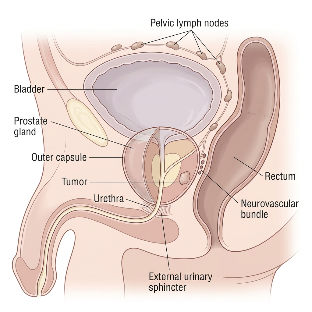
It sits below the bladder, in front of the rectum, and right next to the nerves that control erections. It surrounds the urethra (the tube urine passes through), and lies just above the external urinary sphincter (the muscle that keeps you continent).
This is exactly why treatment decisions are so personal to your body.
- Prostate Gland: The walnut-sized gland itself, sitting directly below the bladder and wrapped completely around the urethra. A tough outer layer called the capsule normally keeps cancer cells contained inside it. Your risk group is really a measure of how likely cells are to have broken through that capsule.
- Bladder: Sits directly above the prostate and stores urine until you are ready to release it. The neck of the bladder connects straight into the top of the prostate, which is part of why bladder and prostate issues are so closely linked.
- Urethra: The tube that carries urine, and semen, out of the body. It runs straight through the center of the prostate on its way down from the bladder, which is exactly why any treatment that disturbs the prostate can also affect urination.
- External Urinary Sphincter: The muscle just below the prostate that you consciously squeeze to stop the flow of urine. It is what keeps you continent, and it sits close enough to the prostate that surgery or radiation can affect it. This is why incontinence is a real risk worth understanding before you choose a treatment and why it is so important to protect this structure when treating the prostate.
- Neurovascular Bundle: The bundle of nerves and blood vessels that control erections. It runs along the outside of the prostate capsule, tucked into the narrow space between the prostate and the rectum. Whether a treatment spares or damages this bundle makes a big difference for preservation of erectile function afterward.
- Rectum: Sits directly behind the prostate, separated by only a thin layer of tissue. That closeness is why radiation can sometimes cause rectal irritation or bleeding, and why surgeons have to work so carefully in such a tight space.
What Is Prostate Cancer?
Prostate cancer starts in the cells of the prostate. Most prostate cancers grow slowly, but some grow fast and need quick action. The job of every Data G is to know which one you have.
The most common type is called adenocarcinoma, which grows from the glandular cells inside the prostate. More than 95 percent of prostate cancers are this type. There are much rarer types like small cell carcinoma, neuroendocrine carcinoma, and ductal carcinoma, which behave differently and need a different game plan.
Prostate cancer is the most common cancer in men in the United States after skin cancer. When it is caught early and the data is read well, outcomes are excellent. When it spreads outside the prostate, the picture changes. That is why early data, smart monitoring, and personalized treatment matter so much.
When it has already traveled outside the prostate, which is the situation this guide is written for, the plan and urgency changes, but that does not mean the outlook is bleak. Smart monitoring and a personalized plan matter more than ever at this point to give yourself every advantage.
Why Does the Capsule Matter?
The prostate is wrapped in a tough outer layer called the capsule. Think of it like the skin of an orange holding everything inside. As long as cancer cells stay inside that capsule, the disease is considered contained.
Sometimes cells break through. They can grow directly through the capsule wall, travel along tiny nerve channels that run into the gland, or slip into nearby lymph vessels. Once they escape, there is a fairly predictable path: first into the tissue just outside the prostate, then to nearby lymph nodes, and if they are not caught there, eventually into the bloodstream, most often settling in bone. This is the path that leads to a metastatic diagnosis.
Understanding this path matters because it explains why your doctors track certain numbers so closely. Your original risk group, your imaging, and your PSA trend are all different ways of asking the same question: how far along this path have the cells traveled?
The Different Types of Prostate Cancer
- Acinar adenocarcinoma: The most common type by far. Starts in the gland cells of the prostate.
- Ductal adenocarcinoma: Rarer and more aggressive than acinar. Often comes with a higher Gleason grade. Worth knowing if this is the type found on your pathology report.
- Neuroendocrine and small cell carcinoma: Rare and aggressive. These types often do not raise PSA the way typical prostate cancer does, so they need a different monitoring and treatment strategy. This type can also show up later as a way the disease adapts after long-term hormone therapy.
- Intraductal carcinoma: A high-risk feature sometimes found on biopsy. It is sometimes linked to BRCA2 mutations and tends to represent more aggressive disease. Ask your doctor whether this was noted on your pathology report.
The Numbers Behind Your Diagnosis

Three numbers come up again and again in prostate cancer, and understanding them helps you follow every conversation with your team.
- Gleason score and Grade Group: It tells you how abnormal the cells look and how urgently they need to be addressed.
- PSA (prostate-specific antigen): A blood test that measures a protein made by prostate cells.
- T-stage: Describes how far the disease had grown at the point it was found, from confined inside the prostate in an early T-stage, to having spread into nearby structures or beyond in a later stage.
Doctors also describe prostate cancer using an overall stage. Stage I and II mean the cancer is still inside the prostate. Stage III means it has grown into nearby tissue like the seminal vesicles. Stage IV, sometimes called metastatic disease, means it has reached lymph nodes, bones, or another organ.
If you were given a Stage 4 diagnosis, or if you find yourself here after an earlier stage came back, this guide is written entirely for you.
PSA Value
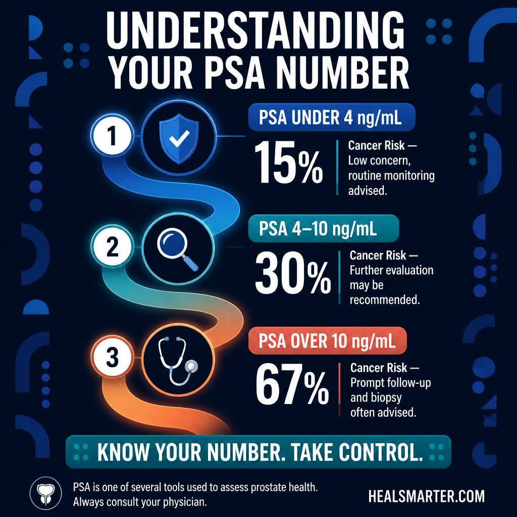
Population guidelines put general ranges at 2.5 to 4.0 ng/mL depending on age, but these are averages built from large groups. Your number needs to be read in the context of your age, your prostate size, and your family history.
| One number alone tells a limited story. A PSA of 5 in a 70-year-old with a large prostate may be less concerning than a PSA of 3 in a 45-year-old with a small gland. Context is everything. |

Gleason Score –
A grading system that describes how normal or abnormal the cells from your biopsy look under a microscope. Cells that look almost normal get a low score. Cells that look very abnormal get a high score. This is one of the most important numbers in this whole journey, and it drives nearly every conversation that follows a positive biopsy.
T Stage

Describes whether an abnormality can be felt by your doctor during a digital rectal exam, and if so, how much of the gland it seems to involve.
Along with your PSA and Gleason score, this is one of the three numbers used to estimate whether the disease is still contained inside the prostate.
PSA Velocity (PSAV): The Speed of Change
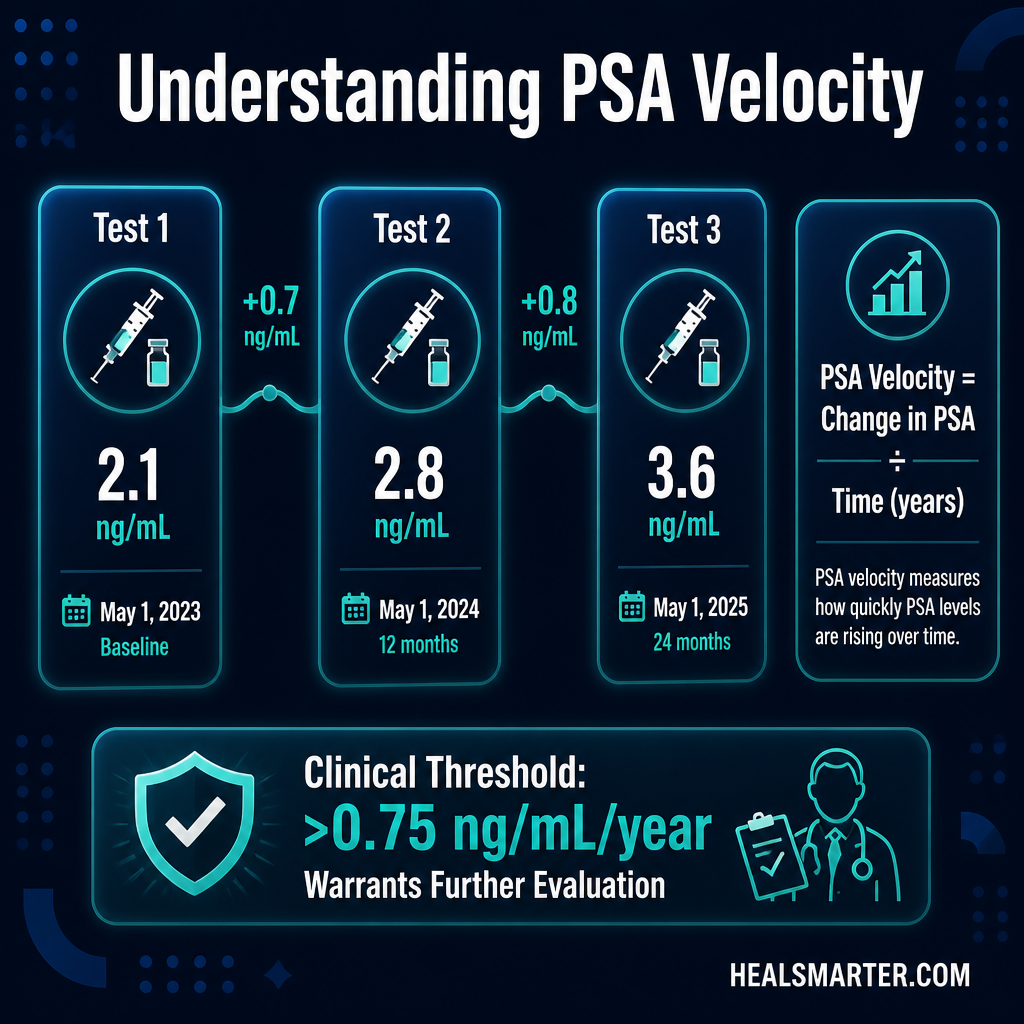
PSA velocity is how quickly your PSA is rising over time.
A PSA that moves from 2 to 4 to 8 in two years tells a very different story than one that moved from 3.5 to 4.0 over five years.
If you only have one PSA number, that may be a problem. You need a trend. Ask your doctor what your previous readings were.
| Ask your doctor: What was my PSA one year ago? Two years ago? Five years ago? If they do not have that data, request your records. Your trend is your most valuable data point. |
PSA Doubling Time
PSA doubling time is how long it takes for your PSA to double. A slow doubling time measured in years suggests a less urgent process. A rapid doubling time measured in months warrants more attention. Free online calculators let you input your own numbers.
Free PSA vs. Total PSA Ratio

PSA exists in two forms in the blood. Free PSA is not bound to proteins. A lower percentage of free PSA relative to total PSA tends to be linked to a higher likelihood of cancer being present. A higher percentage suggests a benign cause like an enlarged prostate. Free PSA value is historically most useful for patients whose PSA is 4-10.
| Ask your doctor: Can we add a free PSA to total PSA ratio to my next blood draw? It is a simple add-on to the same sample that gives you meaningfully more information. |
PSA Density (PSAD)
Your PSA value divided by the size, or volume, of your prostate, usually measured on an MRI or ultrasound. A larger prostate naturally makes more PSA, so density adjusts for that. A high density suggests the elevated number is less likely to simply be a big gland, and more worth a closer look.
Ask your doctor: Can we calculate my PSA density using my MRI prostate volume? It is usually just a quick calculation once both numbers exist.
proPSA (p2PSA) –
A more prostate cancer specific fragment of free PSA. On its own it adds useful detail, and it is also one of three numbers combined into a single score called the Prostate Health Index (PHI), along with total PSA and free PSA. PHI is FDA-cleared and, like the 4Kscore, is an optional tool some men use to get an extra data point before deciding on a biopsy.
Testosterone Value –
Testosterone matters too. Prostate cells are fueled by testosterone, so it is part of the same story as PSA. Think of it this way: PSA is what the prostate produces, and testosterone is what powers it to keep producing. Knowing your baseline testosterone helps your team read your PSA in context, and it becomes essential later if hormone therapy is ever part of your plan.
mpMRI (multiparametric MRI): Now Category 1 evidence as the standard of care before biopsy per NCCN guidelines. It helps target the biopsy and reduces unnecessary procedures.
PSMA expression: Most prostate cancers express PSMA on their cell surface. This drives both PSMA PET imaging and PSMA-targeted radioligand therapy eligibility. (only for intermediate or high risk cancer, not required if PSA is less than 10)
HRR gene status (BRCA2, ATM, CHEK2, BRCA1, and others): Opens the door to PARP inhibitors. BRCA2 is linked to more aggressive disease. Germline testing is now strongly recommended for any high-risk, regional, or metastatic prostate cancer.
MSI-H or dMMR: Rare in prostate cancer (about 3 to 5 percent), but when present opens the door to immunotherapy with pembrolizumab.
| A diagnosis is data. It tells you where you are starting. It does not tell you where you will finish. You are a Data G, and Data G’s use every piece of information to move forward making an informed decision. |
What Metastatic Prostate Cancer Means
Metastatic means cancer cells have broken away from where they started and traveled, through the bloodstream or the lymphatic system, to a distant part of the body. For prostate cancer, that is most often bone, sometimes lymph nodes, and less often the liver or lungs. This is generally considered stage 4 disease.
Here is what matters most for you to understand right now: metastatic does not mean untreatable, and for most men it does not mean an immediate crisis. For example, if only the lymph nodes are involved, there are different schools of thought on if the disease can be eradicated with local therapy alone.
Modern treatment can control this disease for years, often for decades, and the field is moving quickly. Your job, starting today, is to understand exactly which version of this disease you are dealing with, because the right plan depends entirely on the specifics.
The Six Clinical Scenarios
Metastatic prostate cancer is not one situation. It is at least six, and knowing which one applies to you shapes everything that follows.
- Newly diagnosed, low-volume (oligometastatic): Metastatic disease was found the very first time you were diagnosed, with a limited number of spots, usually fewer than four bone spots, none outside the spine or pelvis, and nothing in an organ. This group typically does best with doublet therapy, hormone therapy plus one additional drug, plus radiation aimed at the prostate itself, which has been shown to improve how long the disease stays controlled.
- Newly diagnosed, high-volume: Also found at first diagnosis, but more widespread: four or more bone spots with at least one outside the spine or pelvis, or any spread to an organ. This group usually needs triplet therapy right away, hormone therapy plus chemotherapy plus a second hormone-blocking drug, because more aggressive disease benefits from being met from multiple directions at once.
- Recurrent, low-volume (metachronous oligometastatic): You were treated for what looked like contained disease with surgery or radiation, and a limited recurrence showed up later, usually caught early through a PSMA PET scan. Doctors are increasingly using targeted radiation aimed just at the new spots to delay the need for full-body hormone therapy in this group.
- Recurrent, high-volume: Same story, prior treatment followed by a later recurrence, but this time it is widespread. This moves you into systemic treatment territory, doublet or triplet therapy, depending on your fitness and how much disease is present.
- Metastatic castration-resistant (mCRPC): The disease is growing despite your testosterone being at a castrate, very low, level from ongoing hormone therapy. This does not mean hormone therapy failed completely. It means the cancer cells have adapted, and it is time to add tools: newer hormone-blocking drugs, chemotherapy, PARP inhibitors, radioisotope therapy, or immunotherapy, depending on your biology.
- Node-positive disease: Technically stage 4, but its own separate category. Some doctors believe this can still be cured with local treatment, surgery or radiation, plus hormone therapy. Others believe hormone therapy alone is the safer path. This is genuinely unsettled among experts, which makes it one of the clearest examples of why a second opinion matters.
Where the Disease Has Spread Matters Too
- Bone-only: The most common place for prostate cancer to travel. This carries its own protective steps and on its own tends to carry a more favorable outlook than organ spread.
- Visceral (organ) spread: Spread to the liver or lungs is more aggressive and usually calls for more intensive treatment right away.
- Nodal-only: Spread limited to lymph nodes carries the most favorable outlook of the group.
If This Started as a Rising PSA After Prior Treatment: Your Second Chance Playbook
If you were treated earlier with surgery or radiation and your PSA later rose, the first question your team asked was where the disease came back, because it changes everything that follows:
- Local-only failure: The recurrence is confined to the prostate bed (after surgery) or the prostate gland itself (after radiation), with nothing seen elsewhere on imaging.
- Distant-only failure: No local recurrence, but the disease shows up somewhere else: bone, lymph nodes, or an organ. This calls for systemic treatment.
- Combined local and distant failure: Both at once. This calls for a coordinated plan that usually leads with systemic treatment.
Doctors have a word for treatment aimed at disease that returns after a first attempt at cure. The word is salvage. It is a cold word and you deserve better than it, so we are going to call this what it actually is: your second “at bat” or chance.
A rising PSA after surgery or radiation does not mean you are out of options. For many men it means the opposite. It means something was caught early enough to act on, and there is a real, well-mapped playbook for what comes next. This section walks you through it in the order it will happen.
Step One: Is This Actually a Recurrence?
Before anyone reaches for a plan, the number itself has to hold up. What counts as a recurrence depends entirely on what treatment came first.
- After surgery to remove the prostate: PSA should fall to undetectable. A confirmed PSA of 0.2 ng/mL or higher, measured on two separate tests, is what doctors define as a biochemical recurrence.
- A very low, stable number after surgery: Ultrasensitive tests can pick up tiny amounts of PSA below 0.2 that do not budge over time. This often reflects a small amount of normal prostate tissue left behind, not disease. Guidelines suggest watching this closely rather than rushing into treatment.
- After radiation: The prostate is still in place, so PSA never drops to zero. The definition, called the Phoenix criteria, is a rise of 2.0 ng/mL or more above the lowest point PSA reached. That lowest point is called the nadir.
- After radiation, slow is normal: PSA can take 33 to 44 months to reach its lowest point. A number that is still drifting down two years later is doing exactly what it should.
Read This Before You Panic: The PSA Bounce
If you had radiation and your PSA ticks up somewhere between 12 and 18 months later, stop and breathe. Between 20 and 30 percent of men experience what is called a PSA bounce. It is a temporary, harmless rise that comes back down on its own. It is not a recurrence and it is not a failure.
The way to tell the difference is time. A repeat test in 3 to 6 months usually settles it. Ask your radiation oncologist directly whether your rise fits the bounce pattern before anyone moves you into a treatment decision.
Step Two: How Fast Is It Moving?
One number matters more than the PSA itself, and that is how quickly it is doubling. Your team calculates PSA doubling time by tracking values every 3 to 4 months.
A doubling time under 10 to 12 months signals a recurrence more likely to have traveled, and it usually pushes toward faster action. A slower doubling time buys you room to think. Ask your doctor: what is my PSA doubling time, and how is that shaping what you are recommending?
Step Three: When Do You Get Scanned, and Which Scan?
- Below 0.5 ng/mL: Most guidelines do not recommend imaging yet, because scans are unlikely to show anything at this level.
- Between 0.5 and 10 ng/mL: This is where PSMA PET earns its place. It finds tiny deposits in lymph nodes and bone that standard CT and bone scans miss completely.
- Above 10 ng/mL, or if you have symptoms: Conventional bone scan and CT still have a role and are often used alongside PSMA PET.
- MRI of the prostate or prostate bed: A multiparametric MRI maps whether the recurrence is sitting locally, and it guides a biopsy needle if one is needed.
- Other PET tracers: Fluciclovine (Axumin) and C-11 choline are alternatives, though they are less sensitive than PSMA PET when PSA is very low. Sodium fluoride PET is very sensitive for bone but can light up from arthritis and inflammation, which produces false alarms.
Step Four: Do You Need Another Biopsy?
- After radiation or brachytherapy: A confirmatory biopsy is required before any local treatment aimed at the prostate. These procedures carry real side effects and no one should accept them on imaging alone. Timing matters: the biopsy should not be done sooner than 18 to 24 months after radiation, because cells damaged by radiation die slowly and can look active before then.
- After surgery: A biopsy of the prostate bed is generally not recommended when the physical exam and the MRI are both normal.
Step Five: Your Options, Based on What Was Done First
This is where the playbook splits. A man treated first with surgery gets a very different menu than a man treated first with radiation.
If your first treatment was surgery:
- Radiation to the prostate bed: The standard second-chance treatment when the recurrence looks confined to the pelvis. The dose should be at least 66 Gy. It works best when started early, while PSA is still below 0.5.
- Adding hormone therapy: A 6-month course added to radiation significantly improved how long men stayed free of progression in the GETUG-AFU 16 trial. Higher risk men may be offered up to 24 months, which improves outcomes but carries more side effects. That is a real trade-off and it deserves to be said out loud.
- Adding pelvic lymph node radiation: The SPPORT trial (RTOG 0534) showed that adding radiation to the pelvic lymph nodes plus short-term hormone therapy improved 5-year progression-free survival. Ask whether you are a candidate.
- Surgery to remove affected lymph nodes: An option in select cases with an isolated node found on PSMA PET, usually to delay hormone therapy. Standard practice still leans toward radiation or systemic treatment, because microscopic spread elsewhere is common.
If your first treatment was radiation or brachytherapy:
These options aim at the prostate itself. All of them are harder than they would have been the first time, because radiation leaves scar tissue behind. Every one of them deserves an honest conversation about function before you consent to anything.
- Surgery after radiation: Offers strong local control but is technically demanding. Reported rates of long-term urinary incontinence range from 21 to 93 percent, and erectile dysfunction from 28 to 100 percent. Those are enormous ranges, and surgeon experience is a large part of why. Ask your surgeon for their own numbers, not the numbers in the literature.
- Cryotherapy: Freezing rather than cutting. Less invasive, with lower risk of incontinence and rectal injury than surgery. Often preferred for men over 70 or those with other health conditions. A urethral warming device is essential.
- Brachytherapy as a second treatment: The RTOG 0526 trial reported that 68 percent of men were free from biochemical failure at 5 years, with serious late urinary side effects in about 16 percent.
- HIFU, or focused ultrasound: Used in Europe. In the United States it is not explicitly approved for prostate cancer treatment and the evidence is thinner. Go in with clear eyes.
- Active monitoring: A completely legitimate choice, especially for older men with slow doubling times and other health conditions. Some teams hold off on scans and hormone therapy in men without symptoms until PSA rises above 10.
If your first treatment was focal or partial-gland therapy:
A recurrence can appear in the treated zone, in the untouched part of the gland, or somewhere else entirely. You need an MRI and a biopsy that samples both the treated and the untreated zones, plus a PSMA PET to look further out. Options include repeating the focal treatment, moving to whole-gland treatment, or systemic therapy if disease has traveled.
If your first treatment was hormone therapy alone:
A PSA that rises while you are on hormone therapy is a different situation entirely. It means the disease has become castration-resistant. The very first step is confirming that testosterone is truly at castrate level, because sometimes the real problem is that the hormone therapy is not doing its job. Hormone therapy continues in the background while your team adds tools: an ARPI, chemotherapy, a PARP inhibitor if you carry an HRR mutation, radioligand therapy, or immunotherapy for the small MSI-H group.
Questions to Bring to This Conversation
- Does my PSA rise meet the actual definition of recurrence for the treatment I had?
- Could this be a PSA bounce, and how will we tell?
- What is my PSA doubling time?
- Is a PSMA PET appropriate at my current PSA level?
- Do I need a biopsy before we decide, and has enough time passed since radiation?
- Is this recurrence local, distant, or both?
- What are your personal outcome numbers for the procedure you are recommending?
- If we add hormone therapy, for how long, and why that length?
- What happens to my continence and my sexual function with each option?
- What happens if we watch and wait instead?
A rising PSA is a signal, not a verdict. Get the number, get the doubling time, get the right scan, and then decide.
| A diagnosis is data. It tells you where you are starting. It does not tell you where you will finish. You are a Data G, and Data G’s use every piece of information to move forward making an informed decision. |